
3D model with fluoroscopy (heart imaging)
01 March 2020
Potential of fusion imaging and automated three-dimensional cardiac segmentation during transcatheter aortic valve replacement.
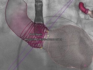
3D model aortic root and left ventricle with fluoroscopy.
Cardiac transcatheter aortic valve replacement (TAVR) is the preferred procedure for aortic valve replacement.
Preprocedural multidetector computed tomography (MDCT) for aortic root analysis and valve prosthesis sizing is considered the standard of care.
However, the use of an iodinated contrast agent during MDCT angiography carries a significant nephrotoxicity risk in patients with chronic severe renal failure. Three-dimensional (3D) transesophageal echocardiographic (TEE) imaging is considered a valuable alternative for aortic root size in this population.
- – – –
(Published: December 2020)(PDF)
Patric Biaggi, Dominik F. Sager, Jeremy Külling, Silke Küest, Christophe Wyss, David Hürlimann, Ivano Reho, Ines Bühler, Georg Noll, Maurus Huber, Oliver Gaemperli, Peter M. Wenaweser, Roberto Corti
Source: Published on behalf of the American Society of Echocardiography. All rights reserved.
More reports

Symposium 2022: Heart valve therapy
Invitation and programme: "Modern Heart Valve Therapy" Dear Ladies and Gentlemen, Dear Colleagues, On Thursday, 2 June 2022, from...

Image fusion in the percutaneous edge-to-edge procedure
Is image fusion necessary or too complex in the percutaneous edge-to-edge repair procedure? Doctor Covadonga Fernandez-Golfin (University Hospital, Madrid, Spain), Prof. Roberto...
The 10 Commandments for the Heart
Heart specialist Prof. Wyss recommends service care from the age of 40 At the latest, every heart needs a service from the age of 40. Cardiologist Prof. Dr....

Cardiac Medicine Hospitals Schaffhausen
Schaffhausen Hospitals join forces with Hirslanden in cardiac medicine Media release dated 17.02.2021 From March 2021, Schaffhausen Hospitals will be working...
Heart medicine at the highest level
The Herzklinik Hirslanden has internationally renowned cardiac specialists with many years of experience in the treatment of heart disease. Thanks to state-of-the-art diagnostics and...
Cardiac Imaging (Heart Imaging)
Techniques of imaging the heart (cardiac imaging): Imaging the heart (cardiac imaging) is indispensable for making a diagnosis....
All specialists at the Heart Valve Center of Herzklinik Hirslanden
Prof. Dr. med. ROBERTO CORTI
Interventional Cardiology
Prof. Dr. med. JÜRG GRÜNENFELDER
Cardiac Surgery
MD. THIERRY AYMARD
Cardiac Surgery
PD Dr. med. PATRIC BIAGGI
Cardiology | Imaging
Prof. Dr. med. OLIVER GÄMPERLI
Interventional Cardiology
PD Dr. med. DAVID HÜRLIMANN
Cardiology | Rhythmology
Dr. med. IOANNIS KAPOS
Cardiology | Imaging
MD. SILKE WÖRNER
Cardiology | Imaging
Prof. Dr. med. GEORG NOLL
Cardiology | Prevention
MD. IVANO REHO
Cardiology | Aortic Aneurysm
PD Dr. med. (H) DIANA RESER
Cardiac Surgery
Prof. Dr. med. JAN STEFFEL
Cardiology | Rhythmology
Prof. Dr. med. PETER M. WENAWESER
Interventional Cardiology
Prof. Dr. med. CHRISTOPHE WYSS
Interventional Cardiology