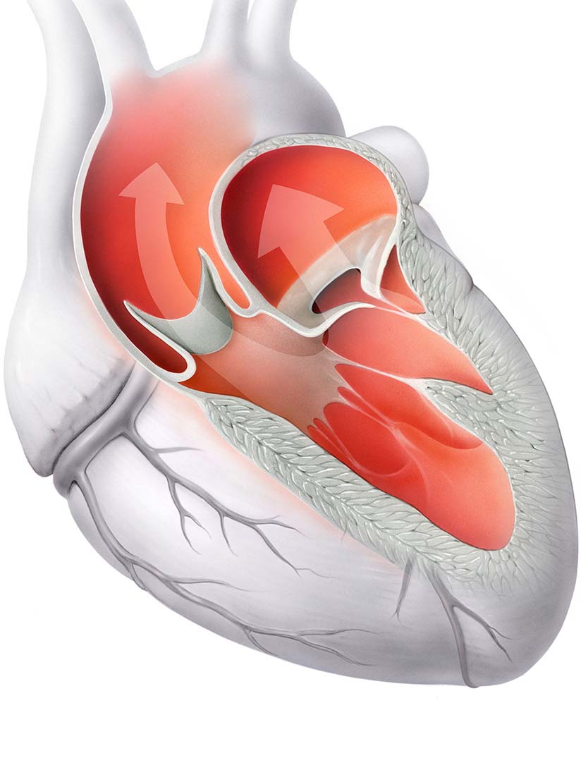
Function of the healthy heart valves
Each half of the heart has a sail flap and a pocket flap.
The four heart valves act as valves in the heart to ensure regulated blood flow throughout the body. The heart valves are located between the atria and the ventricles, and between the ventricles and the outgoing arteries (aorta and pulmonary artery).
Function of the healthy heart valves
The four heart valves are the valves in the heart that ensure the orderly flow of blood in the heart. They are formed by the inner lining of the heart (endocardium). The valves control the flow of blood from the atria to the ventricles and from the ventricles to the great arteries by opening and closing in a timed manner.
The tricuspid and mitral valves are located between the atria and the ventricles. The pulmonary valve and aortic valve are located at the junction of the ventricles and the large blood vessels, the pulmonary artery and the aorta, respectively.
The Mitral valve consists of two leaflets. The leaflets of the mitral valve are attached to the papillary muscles facing the ventricle by tendon threads.
The Tricuspid valve consists of three connective tissue leaflets. The leaflets of the tricuspid valve are attached to the papillary muscles facing the ventricle by tendon threads.
The Aortic valve consists of three flap pockets. The pockets are indented towards the heart and can fill with blood. The aortic valve is slightly larger than the related pulmonary valve because of the greater stress placed on it.
Like the aortic valve, the Pulmonary valve The pulmonary valve is made up of three crescent-shaped pockets (semilunar valve). The pulmonary valve is much less frequently affected by valve diseases.
Diseases and malformations of the heart valves can occur when there is a narrowing (stenosis) of a heart valve so that it blocks the flow of blood. Or a heart valve leaks (insufficiency), allowing blood to flow back into the heart. Sometimes a heart valve has both stenosis and insufficiency.

Anatomy of the heart valves
The pulmonary valve and Aortic Valve (left heart valve in the picture) are located at the junction of the chambers with the large blood vessels, the pulmonary artery and the coronary artery, respectively.
The tricuspid valve and Mitral Valve (right heart valve in the picture) are located between the atria and the ventricles.
The following diseases of the heart valves are treated at the Heart Valve Center:
Specialists at the Heart Valve Center
Do you have questions about the function of healthy heart valves?
Make an appointment with the specialists at the Heart Valve Center.
 The heart is a muscular hollow organ that pumps blood through the body with rhythmic contractions and in this way ensures the supply of all organs.


